If your cardiologist has recommended a cardiac PET/CT scan, you might have questions about what the test involves and why it is necessary. This guide will walk you through the technology, the process, and what it reveals about your heart health.
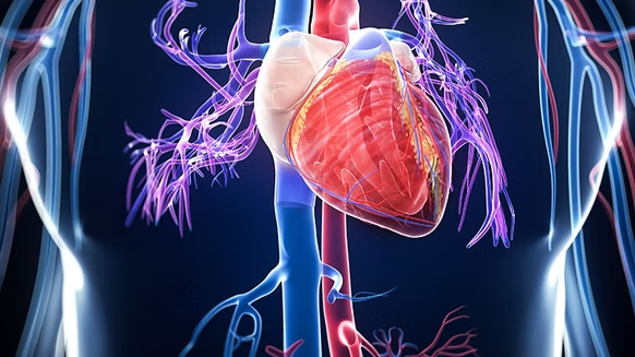
Cardiac PET/CT is a hybrid imaging technique that combines two advanced imaging modalities—PET (Positron Emission Tomography) and CT (Computed Tomography)—into a single, comprehensive test. The result is the most accurate, detailed assessment of your heart blood flow and function available today.
What Is PET/CT Imaging?
PET/CT imaging fuses two complementary technologies:
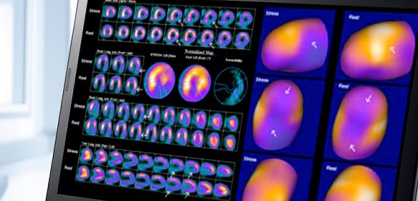
PET (Positron Emission Tomography) creates a functional image—it shows how your heart is working by tracking blood flow and metabolic activity. A small amount of radioactive tracer is injected into your bloodstream. This tracer accumulates in areas where blood flow and metabolism are active. A specialized camera detects the tracer signals and creates detailed images of perfusion (blood flow) throughout your heart muscle.
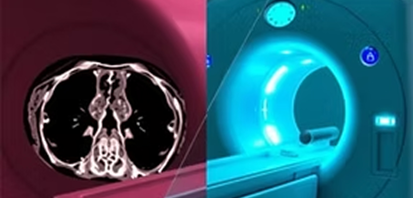
CT (Computed Tomography) creates a structural image—it shows what your heart looks like. X-rays are used to create detailed cross-sectional images of your heart and coronary arteries, revealing any calcifications, blockages, or anatomical abnormalities.
By fusing these two images together, your cardiologist sees both function and structure in precise anatomical detail. This hybrid approach is far more powerful than either test alone.
Why Is PET/CT Imaging Important for Cardiac Patients?
- Coronary Artery Disease (CAD) Diagnosis — PET/CT directly visualizes blood flow to your heart muscle, identifying blockages in coronary arteries with exceptional sensitivity and specificity. Unlike stress tests that measure indirect signs, PET/CT shows your doctor exactly where blood flow is compromised.
- Myocardial Perfusion Assessment — The PET portion reveals how well oxygen-rich blood is reaching every region of your heart muscle, both at rest and during stress. Areas with reduced perfusion indicate ischemia (insufficient blood flow) or prior damage.
- Myocardial Viability Assessment — For patients with previous heart attacks or reduced heart function, PET/CT can determine which heart tissue is still viable and could improve with revascularization, and which is permanently damaged. This is crucial for surgical planning.
- Comprehensive Risk Stratification — The combination of detailed perfusion imaging and CT coronary calcium scoring allows for precise risk assessment and guides treatment decisions.
How Does Cardiac PET/CT Imaging Work?
Understanding the process can help ease any concerns about the test:
Preparation
You will receive specific instructions before your scan. Typically, you should avoid caffeine for 24 hours (as it can affect test results) and eat only a light meal a few hours before arrival. Wear comfortable clothing without metal (snaps, zippers, or underwire). Plan for the appointment to last about 3-4 hours.
PET Imaging
A small amount of radioactive tracer (typically rubidium-82 or ammonia-13 for cardiac studies) is injected into your arm through an IV. This is a very small dose—it is safe and leaves your body naturally within hours. The tracer travels through your bloodstream to your heart muscle.
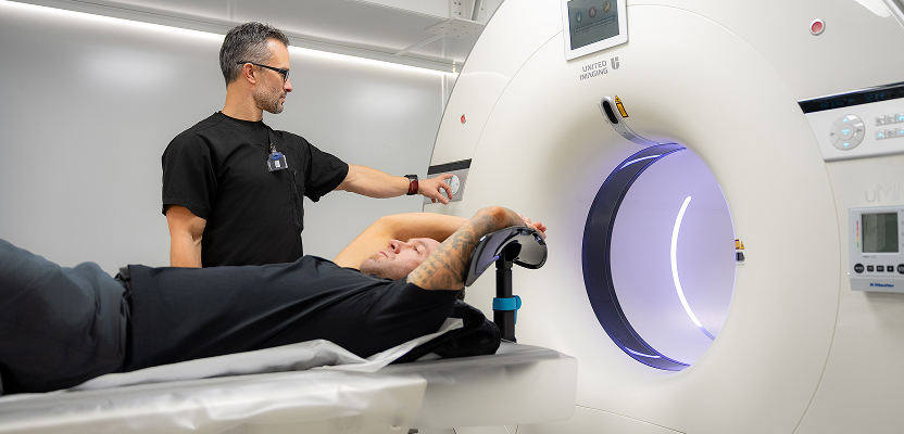
You will lie on a scanning table while a PET camera (a large ring-shaped device) rotates around your chest, detecting the tracer signals and creating detailed images of blood flow. You may undergo imaging at rest first, then again during cardiac stress (either from treadmill exercise or IV medication that simulates exercise stress).
CT Imaging
After the PET imaging, you will have a CT scan. You will lie still on the scanning table while an X-ray tube rotates around your chest, taking many detailed cross-sectional images. This scan is brief—usually only a few seconds of actual imaging. The CT provides detailed anatomical information and allows for calcium scoring of your coronary arteries.
Image Fusion
A computer fuses the PET and CT images together, creating three-dimensional representations that show blood flow overlaid on anatomical structure. Your cardiologist reviews these fused images to identify any abnormalities.
Benefits of PET/CT Imaging for Cardiac Patients
- Early Detection — PET/CT can identify coronary artery disease and myocardial ischemia before you experience a heart attack. This early detection allows for preventive treatment.
- Superior Accuracy — PET/CT has higher sensitivity and specificity for detecting coronary artery disease compared to traditional stress testing or other imaging modalities.
- Non-Invasive Assessment — Unlike cardiac catheterization (which requires insertion of a catheter into blood vessels), PET/CT provides detailed information without invasive procedures.
- Precision Medicine — The detailed information guides treatment decisions: who needs medication, who needs intervention, and who can be managed with lifestyle changes.
- Treatment Monitoring — PET/CT can assess how well treatments are working and monitor changes in your heart function over time.
PET Versus SPECT: Understanding the Difference
You may hear about both PET and SPECT (Single Photon Emission Computed Tomography) imaging for cardiac assessment. While both are nuclear cardiac imaging techniques, PET offers important advantages:
- PET imaging uses positron-emitting tracers that provide superior image quality, better spatial resolution, and faster acquisition times. The technology provides more accurate quantification of blood flow and is particularly valuable for complex cases.
- SPECT imaging uses single photon-emitting tracers. While SPECT is effective and more widely available, it generally provides lower image quality and may require longer imaging times or have limitations in obese patients.
For the most accurate cardiac assessment, PET/CT is the gold standard. At VIP Imaging, we specialize in mobile cardiac PET/CT services, bringing this advanced technology to Southern California patients.
What to Expect During Your PET/CT Scan
Here is a practical walkthrough of what a typical patient experiences:
- Arrival and Check-In — You will check in 10-15 minutes early. The staff will review your medical history, answer questions, and verify you have followed pre-scan instructions.
- IV Line Placement — A nurse will place an IV line in your arm. This is small and usually painless. The IV is used to inject the tracer and, if needed, medications for stress testing.
- Initial Imaging — You will lie on the scanning table with electrodes placed on your chest (these monitor your heart rhythm). The PET camera will rotate around you for about 15-20 minutes while imaging at rest.
- Stress Phase — If a stress component is needed, you will exercise on a treadmill while monitored, or you will receive IV medication that increases your heart rate. Stress imaging then occurs.
- CT Imaging — After PET imaging, you will have your CT scan. You will need to hold your breath briefly (usually 10-15 seconds) while the X-ray tube scans your chest.
- Recovery — After scanning is complete, the IV is removed. There are no restrictions—you can return to normal activities immediately. Drink plenty of water to help flush the tracer from your body.
Who Should Consider Cardiac PET/CT Imaging?
Cardiac PET/CT may be appropriate if you have:
Symptoms suggestive of coronary artery disease, such as chest pain, shortness of breath, or unusual fatigue. Significant risk factors for heart disease, including family history, diabetes, hypertension, high cholesterol, or smoking history. A history of previous cardiac events and need assessment of remaining heart tissue viability. Reduced heart function where your doctor wants to determine if revascularization could help. Inconclusive or conflicting results from other cardiac tests.
Your cardiologist can determine if PET/CT is the right test for your specific situation.
The VIP Imaging Difference
At VIP Imaging, we bring state-of-the-art mobile cardiac PET/CT imaging directly to Southern California patients. Rather than traveling to distant imaging centers, our mobile units deliver the same advanced technology found in major medical centers—right in your community.
We understand that cardiac health concerns can be stressful. Our compassionate, experienced team makes the imaging process as comfortable and convenient as possible. We work closely with your cardiologist to ensure you get the precise imaging data needed for confident diagnosis and optimal treatment planning.
VIP Imaging serves patients throughout Southern California with the most advanced cardiac imaging technology available.
Next Steps: Schedule Your Cardiac PET/CT Scan
If your cardiologist has recommended a cardiac PET/CT scan, or if you want to explore whether PET/CT imaging is right for your cardiac health concerns, contact VIP Imaging today.
Let us provide you with the most accurate, detailed cardiac assessment available—conveniently delivered to your area. Our board-certified nuclear cardiologists and experienced imaging technicians are ready to help you take control of your heart health.